


Preparing and observing a microscope slide


Lesson details
Learning outcome
I can prepare a microscope slide with a specimen of tissue, observe it using a microscope, and record my observations in a scientific line drawing.
Key learning points
- The cells that make up tissue can be viewed using a light microscope.
- A sample of tissue can be prepared on a slide to view using a microscope.
- Adding a stain can make it easier to see some parts of cells.
- Changing the magnification and focus of a light microscope enables stained cell structures to be observed.
- Observations from a light microscope can be recorded in a scientific line drawing.
Keywords
Tissue - A tissue is a group of similar cells with the same job working together.
Slide - To observe a specimen using a light microscope, we have to put a thin layer of it on a glass microscope slide.
Stain - A coloured liquid put onto a specimen so that the cells and their structures can be more easily seen with a light microscope.
Scientific line drawing - Observations from a light microscope can be recorded by making a labelled scientific line drawing.
Common misconception
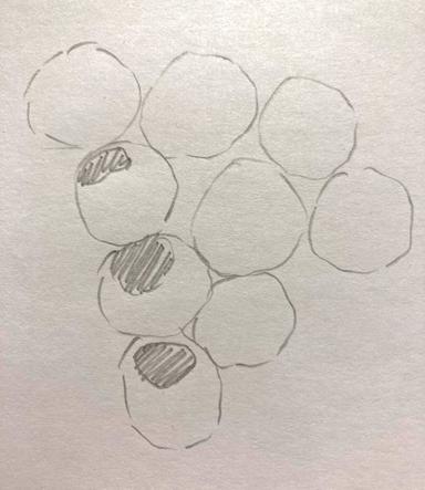
Producing a line drawing with sketched broken lines, shading, no labelling and no magnification.
Learning cycle 2 in this lesson explores how to produce a scientific line drawing to record observations from light microscopy.
Teacher tip
Pupils should be provided with the opportunity to produce their own line drawings to record observations from light microscopy, and perhaps to peer review them in a constructive way.

Content guidance
Risk assessment required - equipment

Supervision
Adult supervision required

Licence

Lesson video
Loading...
Prior knowledge starter quiz
6 Questions
Q1.A is a piece of equipment that is used to see things that are very small.
Q2.Which parts of the microscope magnify an image?
Q3.Which is not a part of a microscope?
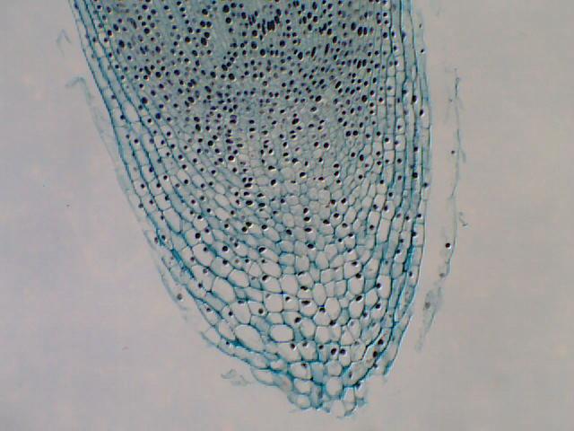
Q4.Which action would you take to increase the magnification of this image (to make the individual cells appear larger)?
Q5.A student set up a microscope with 10x eyepiece and a 40x objective lenses. What is the total magnification?
Q6.A is a piece of curved glass that focuses and magnifies light.
Assessment exit quiz
6 Questions
Q1.Which piece of apparatus is not used to prepare an onion tissue slide?
Q2.Match the apparatus to its job in preparing a microscope slide.

Q3.Starting with a glass slide, put these stages in the correct order for preparing a microscope slide.
Q4.The chemical that is placed on a specimen so that it can been seen easily is called a...
Q5.Which features should be altered in this scientific line drawing to improve it?
Q6.Which of these descriptions is not a key feature of an accurate scientific line drawing?
To help you plan your 7 science lesson on: Preparing and observing a microscope slide, download all teaching resources for free and adapt to suit your pupils' needs...
To help you plan your 7 science lesson on: Preparing and observing a microscope slide, download all teaching resources for free and adapt to suit your pupils' needs.
The starter quiz will activate and check your pupils' prior knowledge, with versions available both with and without answers in PDF format.
We use learning cycles to break down learning into key concepts or ideas linked to the learning outcome. Each learning cycle features explanations with checks for understanding and practice tasks with feedback. All of this is found in our slide decks, ready for you to download and edit. The practice tasks are also available as printable worksheets and some lessons have additional materials with extra material you might need for teaching the lesson.
The assessment exit quiz will test your pupils' understanding of the key learning points.
Our video is a tool for planning, showing how other teachers might teach the lesson, offering helpful tips, modelled explanations and inspiration for your own delivery in the classroom. Plus, you can set it as homework or revision for pupils and keep their learning on track by sharing an online pupil version of this lesson.
Explore more key stage 3 science lessons from the Cells unit, dive into the full secondary science curriculum, or learn more about lesson planning.